

Urine Microscopical Examination
MICROSCOPICAL EXAMINATION OF URINE

Preperation of slide
Morning midstream urine or fresh
random urine 3-4 hours after the patient has last voided is satisfactory
After
2 hour cells & cast begin to lysis.
Refrigeration (2-8) prevent from lysis
Refrigeration (2-8) prevent from lysis
Preparation
of slide -
Take 10-15 ml urineà centrifuge at 2000 rpm x 5min à discard the supernatants --> take a drop of deposit on slide -> cover with 18mm squire cover slip.
Then see under microscope in high power, condrnser down , iris small setting.
Urine should be examined within one hour after collection. otherwise preservative should be used (Formalin/ thymol)
Comment on:
Squamous/
transitional epithelial cell present if large number or as fragments.
No.
Of cell in / HPF
Bacteria/
yeast/ microorganism as (2+ like)
Crystals.
Abnormal crystal should be confirmed chemically.
On
large amount of mucus.
MICROSCOPICAL FINDINGS TO NOTE:
MICROSCOPICAL FINDINGS TO NOTE:
Organised sediments
RBC
WBC
Epithelial
cell
Squamous
Transitional
Renal
tubular
Casts
Matrix casts
Hyaline
cast- formed almost with tamm-Horsfall protein. / upto 2 /HPF is normal. /
found normally in – exercise/ old/ HTN/ CCF/ dehydration/ Fever
Waxy
cast : common in CRF. If broad- called Renal failure cast
Inclusion cast
Granular
cast – protein[ fibrinogen/ Immune complex/ globulin] , cell debris [cell
damage & degeneration]
Fatty
cast : in heavy proteinuria, in normal saline.
Hemosiderin
granules-
Melanin
granules
Pigments cast :
Hemoglobin-
typically yellow/ red. Found with RBC cast in glomerular disease. & in
tubular bleeding/ hemoglobinuria.
Myoglobin
: red brown. Found in myoglobinuria following muscle damage.
Bilirubin
: deep yellow brown cast. Found in obs. Jaundice.
Drug
Cellular cast:
RBC
cast- condition when RBC appear in urine, like- AGN/ SABE/ IgA nephropathy/
Tubulo-interstitial disease.
Leucocytic
cast:- mostly in Tubulo-interstitial disease., inflammation/ glomerular
disease.
Renal
tubular epithelial cell cast : ATN/ viral disease/ drug toxicity/ heavy metal
toxicity.
Mixed
cell
Broad cast :
Dismater 2-6 times than normal cast, due to tubular dilation and/or
stasis in collecting duct. Typically seen in CRF, has poor prognosis. (Renal
failure cast)
Telescoped
sediment : simultaneous finding of elements of GN + NS. Include – RBC, RBC
cast, Cellular Cast, Broad Waxy cast, oval fat bodies, fatty cast, lipid
droplet. Found in – SABE/ collagen vascular disease (Lupus nephritis).
Abnormal cell & other formed elements
Tumor
cell
Viral
inclusion cell
Platelets
Bacteria
Fungi
Spermatozoa
Parasie
Trichomonas vaginalis-
motile found if female, contaminated/ urethral, bladder infection. Schistosomia and E.Histolytica
ova also may be found.


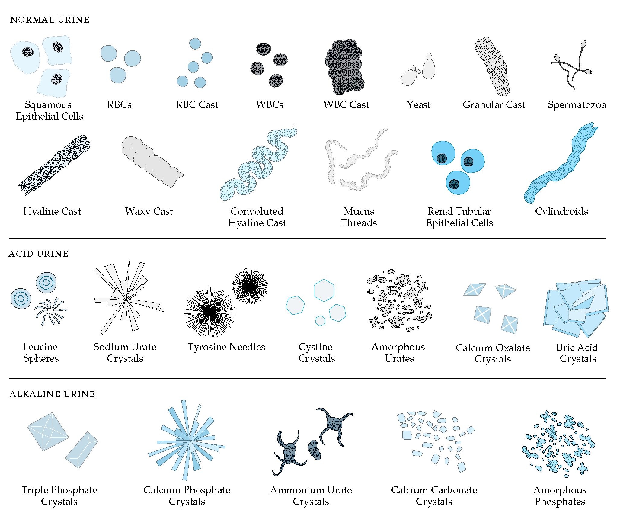
Urine examination findings
Un-organised crystal
Formed by ppt. Of urinary salts when PH, Temperature & concentration of
urine changes. These appear either amorphous or crystalline form. Crystal forms
on standing/ refrigeration, in vivo, main cause is increase of solute
concentration.]
Crystal found in
normal acid urine :
Amorphous urates:
(Calcium, Magnesium, Sodium,
Potassium) : dissolve by heat & NaOH, not by acetic acid.
Crystalline urates
( Sodium, potassium, Ammonium): on addition of acetic acid- turn to
crystal f uric acis, can be seen microscopically .
Crystalline uric acid
4 sided, flat, yellow/ reddish brown, rhombic plates or prism
Calcium oxalates
occurs in Ph-6, nutral, slight alkaline urine], often associated
with spinach,Tomato.
Crystal found in normal alkaline urine
Amorphous phosphates
(Calcium, Magnesium). [ found in
alkaline/ slightly acidic ph] – dissolve in acetic acid.
Crystalline phosphates.
( triple phosphate :
ammonium- magnesium-calcium) : presence of fresh urine indicate stone in
kidney/ bladder.
6
sided prism
Leaf
like
Calcium carbonate
[rare in man.] small colourless sphere/ granules. Soluable in
acetic acid with gas formation.
Ammonium biurate
[ in alkaline/ nutral/ slightly acidic urine]
Usually
found with phosphate crystal.
Yellow/
brown sphere showing concentric striation.
Dissolve
in acetic acid + heat.
Crystal found in abnormal urine
Leucine
Oil drop like with radial & concentric striation.
Tyrosine
fine needle like, called Tyrosine needle.
Cystine
hexagonal,
colorless, plate like.
Found in acidic urine- resemble uric acid
crystal
Found
in cystinuria- congenital renal tubular defect.
Drug crystal
sulphonamide, ampicillin, radiographic material, others.
Artifacts
Air
bubble
Fibre
Oil
Hair


No comments